联系我们
意见反馈

关注公众号

获得最新科研资讯

陈素明课题组(质谱研究组)

简介 化学与生物质谱

分享到

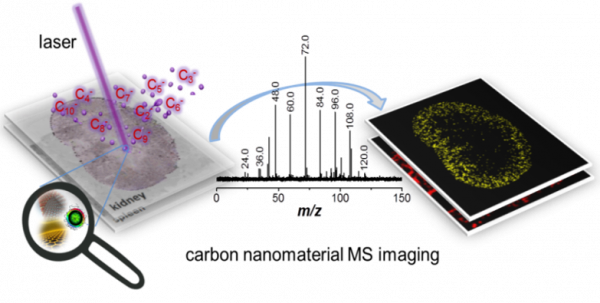
Mass spectrometry imaging reveals the sub-organ distribution of carbon nanomaterials

2015
期刊 Nature Nanotechnology
Label and label-free methods to image carbon-based nanomaterials exist. However, label-based approaches are limited by the risk of tag detachment over time, and label-free spectroscopic methods have slow imaging speeds, weak photoluminescence signals and strong backgrounds. Here, we present a label-free mass spectrometry imaging method to detect carbon nanotubes, graphene oxide and carbon nanodots in mice. The large molecular weights of nanoparticles are difficult to detect using conventional mass spectrometers, but our method overcomes this problem by using the intrinsic carbon cluster fingerprint signal of the nanomaterials. We mapped and quantified the sub-organ distribution of the nanomaterials in mice. Our results showed that most carbon nanotubes and nanodots were found in the outer parenchyma of the kidney, and all three materials were seen in the red pulp of the spleen. The highest concentrations of nanotubes in the spleen were found within the marginal zone.